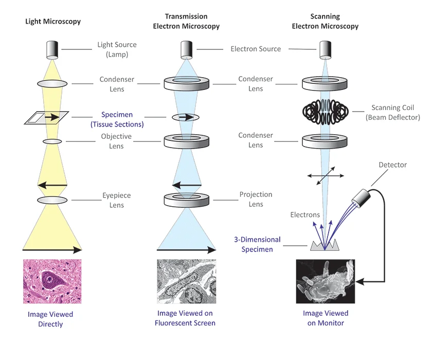
Differences between Light Microscope and Electron Microscope
| Light Microscope | Electron Microscope |
|---|---|
| Illuminating source is the Light. | Illuminating source is the beam of electrons. |
| Specimen preparation takes usually few minutes to hours. | Specimen preparation takes usually takes few days. |
| Live or Dead specimen may be seen. | Only Dead or Dried specimens are seen. |
| Condenser, Objective and eye piece lenses are made up of glasses. | All lenses are electromagnetic. |
| It has low resolving power (0.25µm to 0.3µm). | It has high resolving power (0.001µm), about 250 times higher than light microscope. |
| It has a magnification of of 500X to 1500X. | It has a magnification of 100,000X to 300,000X. |
| The object is 5µm or thicker. | The object is 0.1µm or thinner. |
| Image is Colored. | Image is Black and White. |
| Vacuum is not required. | Vacuum is essential for its operation. |
| There is no need of high voltage electricity. | High voltage electric current is required (50,000 Volts and above). |
| There is no cooling system. | It has a cooling system to take out heat generated by high electric current. |
| Filament is not used. | Tungsten filament is used to produce electrons. |
| Radiation risk is absent. | There is risk of radiation leakage. |
| Specimen is stained by colored dyes. | Specimen is coated with heavy metals in order to reflect electrons. |
| Image is seen by eyes through ocular lens. | Image is received in Zinc Sulphate Fluorescent Screen or Photographic Plate. |
| It is used for the study of detailed gross internal structure. | It is used in the study of external surface, ultra structure of cell and very small organisms. |
 |  |
No comments:
Post a Comment